Advanced Nuclear Medicine Services in Kerela
Delivering accurate diagnostics and targeted therapies with cutting-edge molecular imaging technology.
At DDNMRC, we offer advanced Nuclear Medicine and Molecular Imaging services through our state-of-the-art PET CT and SPECT CT systems. Located near Medical College in Kerala, our center is equipped with the latest diagnostic technologies and led by a highly skilled team of nuclear medicine specialists dedicated to accurate diagnosis and personalized care.
Our goal is to go beyond conventional imaging by offering functional and molecular-level insights that enable precise understanding of diseases at an early stage. With cutting-edge tools such as PET-CT and Gamma Camera systems, we provide detailed information on metabolic activity, organ function, blood flow, and tumor detection—helping clinicians make confident decisions for effective treatment planning.
Nuclear Medicine
Our Vision
We are committed to transforming healthcare through precision diagnostics. By combining structural and functional imaging, our focus is to provide more personalized, patient-specific care. Functional Imaging in Nuclear Medicine allows us to view disease processes in real time—something traditional imaging alone cannot offer. This approach helps shift from generalized care to Personalized Medical Care, improving diagnosis, treatment outcomes, and overall patient experience.
Why Choose DDNMRC
Our center is built on a strong foundation of medical excellence and patient-centric values.
- We house world-class imaging equipment with high-resolution capabilities.
- Our team of experienced nuclear medicine physicians and technologists ensures accurate reporting and compassionate care.
- Each step of our process is designed for maximum comfort and efficiency, following international safety and quality standards.
From early detection to targeted therapy monitoring, DDNMRC continues to be a trusted name in nuclear medicine diagnostics across Kerala and beyond.

Our Technology

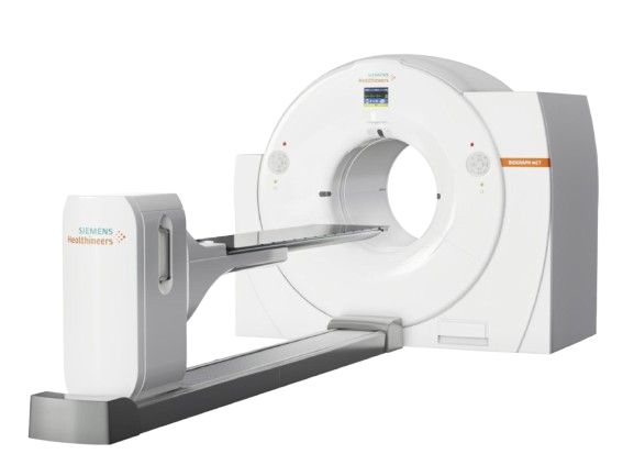
01. Siemens Biograph mCT Flow PET-CT
Our department is equipped with the advanced Siemens Biograph mCT Flow PET-CT scanner, designed to deliver high precision and efficiency. This hybrid imaging system uses Time of Flight (TOF) technology, which enhances PET image clarity, allowing for superior contrast and the detection of even the smallest tumors with accurate anatomical detail. Unlike standard 16-slice systems, this scanner is integrated with a 40-slice CT, enabling high-quality imaging and faster scans. The combination of TOF and advanced CT reduces patient radiation exposure while ensuring excellent image quality and diagnostic accuracy.

02. Siemens Biograph mCT Flow PET-CT
Our department is equipped with the advanced Siemens Biograph mCT Flow PET-CT scanner, designed to deliver high precision and efficiency. This hybrid imaging system uses Time of Flight (TOF) technology, which enhances PET image clarity, allowing for superior contrast and the detection of even the smallest tumors with accurate anatomical detail. Unlike standard 16-slice systems, this scanner is integrated with a 40-slice CT, enabling high-quality imaging and faster scans. The combination of TOF and advanced CT reduces patient radiation exposure while ensuring excellent image quality and diagnostic accuracy.

03. Siemens Biograph mCT Flow PET-CT
Our department is equipped with the advanced Siemens Biograph mCT Flow PET-CT scanner, designed to deliver high precision and efficiency. This hybrid imaging system uses Time of Flight (TOF) technology, which enhances PET image clarity, allowing for superior contrast and the detection of even the smallest tumors with accurate anatomical detail. Unlike standard 16-slice systems, this scanner is integrated with a 40-slice CT, enabling high-quality imaging and faster scans. The combination of TOF and advanced CT reduces patient radiation exposure while ensuring excellent image quality and diagnostic accuracy.

Services
Nuclear medicine studies give valuable information in most of the medical fields:
- Thyroidology / Endocrinology
- Medicine / Surgery
- Oncology
- Rheumatology
- Urology
- Orthopaedics
- Cardiology
- Neurology / Psychiatry
- Paediatrics
- Pulmonology
- Nephrology
- Gastroenterology
- Diabetology
This records the distribution of a radioactive tracer (Technetium-99m or Iodine-i 31) in the thyroid gland
1. Thyroid scan pertechnetate (99m Tc04-)
• Evaluation of congenital hypothyroidism
• Function of palpable thyroid nodule
• Detection of ectopic thyroid tissue
• Evaluation of substemal mass-Midline neck swelling
• Thyromegaly and toxic goiters • Evaluation of thyroiditis
2. Whole body and thyroid scan-Sodium iodide (Na131 |)
• Assessment of remnant thyroid tissues in post-operative thyroid Cancer
• Visualisation of thyroid cancer & metastases in the body
• Follow up evaluation of recurrence
3. Parathyroid Scan-Pertechnetate (99m Tc04-) and 99m Tc-MIBI
• Detection and localization of Parathyroid adenoma
4. 131 I-MIBG Scan
• Detection of adrenal and extra-adrenal pheochromocytoma
• Neuroendocrine tumors (NETs)
5. Radioactive Iodine Uptake Test (RAIU)
RAIU can be determined using either I-131 (7 uCi) or I-123 (200-300 uCi). Normal 24 hour RAIU is between 8 to 35%. Normal 4 hour RAIU is generally between 5 to 15%.
% UPTAKE = ((Neck Counts – Net Thigh Counts) X 100)
(Net Standard Counts)
The RAIU test provides a useful assessment of thyroid function: in general, the higher the iodine uptake, the more active the gland. Note, however, that in patients with hypothyroidism such as Hashimoto’s disease, the % uptake may be low, normal or high depending on the steps affected in thyroid hormone synthesis. This is therefore of no value in establishing the diagnosis of hypothyroidism.
Indications
RAIU test may be helpful in the following clinical conditions:
a) To confirm hyperthyroidism
b) To calculate therapeutic dose of I-131
c) To determine autonomous thyroid tissue (i.e. toxic nodules, – combined with thyroid scan)
d) To determine the cause of thyrotoxicosis*
* The most useful role of RAIU test is in determining the etiology of thyrotoxicosis. Thyrotoxicosis simply refers to excess thyroid hormone in the body and may be due to overactivity of the thyroid gland (hyperthyroidism), or other causes such as inflammation of the gland (thyroiditis) or ingestion of excess thyroid hormone. In “true hperthyroidism” – RAIU uptake will be high while thyrotoxic patients with thyroiditis or who abuse thyroid hormones will have a low RAIU. Other factors that affect RAIU uptake are listed below.
Patient’s iodine pool:
-Dietary variations: RAIU can be falsely elevated in patients who are iodine deficient.
– Renal function: Poor renal function results in decreased excretion of iodine, the larger iodine pool will compete with the radiopharmaceutical for uptake thus falsely lowering RAIU.
– Recent iodine contrast study: The excess iodine will falsely lower the RAIU. It may require weeks prior to scanning in order to obtain an accurate value. The Wolff-Chaikoff effect refers to a transient decrease in iodide organification and hormone synthesis in normal or Graves patients following an iodide load.
Medications
– Antithyroid drugs: Propythiouracil and methimazole result in a poor 24 hour scan because they block iodide oxidation and organification (and to a lesser extent thyroid hormone excretion). Because the agents do not inhibit iodine trapping, a pertechnetate scan, may be technically adequate.
– Thyroid hormone
– Amiodarone
* Hyperthyroidism (Grave’s disease or TSH-secreting pituitary adenoma)
* Rebound following abrupt withdrawal of antithyroid meds
* Long term antithyroid therapy: Prolonged therapy may result in decreased circulating levels of T4- hence TSH levels will increase. This will accentuate radioiodine uptake.
* Enzyme defects
* Iodine starvation
* Lithium Therapy
* Early Hashimoto’s Thyroiditis (Patients rarely present at this stage, RAIU is typically normal [early] or decreased [late] in these patients)
* Rebound during recovery from subacute thyroiditis
* Blocked Trapping:
– Iodine load (most common). An iodine load can “falsely” lower the RAIU through two mechanisms: 1) producing the Wolf-Chaikoff effect 2) causing a dilutional effect, i.e., diluting the I-123 atoms with nonradioactive I-127 atoms.
– Exogenous thyroid hormone replacement depressing TSH levels
– Ectopic thyroid: Struma Ovarii
* Blocked Organification:
– Antithyroid medication (PTU): Note- Tc-99m uptake should not be affected
* Parenchymal Destruction:
– Acute, Subacute and Chronic Lymphocytic Thyroiditis
* Hypothyroidism:
– Primary or Secondary (insufficient pituitary TSH secretion)
– Surgical/Radioiodine Ablation of Thyroid
The thyroid suppression test is based on the premise that normal thyroid activity is suppressed by exogenously administered thyroid hormone. The test is used to determine the autonomy of a hot nodule or diffusely enlarged gland. A baseline scan and uptake are performed. The patient is then placed on T3 (Cytomel 25 ug TID [50-75 ug daily] for 7 days) to suppress TSH production. Iodine uptake should normally fall to 50% of presuppression value in a non-autonomously functioning nodule.
Indications
In most cases of hyperthyroidism, the diagnosis will be apparent on baseline scan and uptake. In Graves’ disease, the uptake will be elevated with a homogenous distribution. In autonomous nodules, uptake may be high normal or elevated with the remainder of the normal gland suppressed. This together with the availability of sensitive TSH assays has virtually eliminated the need to do thyroid suppression test. Some conditions where this test may provide additional information include:
*Early Graves’ disease with borderline hyperthyroidism or in euthyroid Graves’ disease (patients presenting with opthalmopathy but normal function tests).
*Nodules which are indeterminate (warm or nondelinated) where a distinction between hot and cold is unclear. A repeat scan on cytommel (T3) or levothyroxine therapy with suppression of TSH may reveal whether the thyroid nodule is autonomous (hot on non autonomous (cold). A non autonomous nodule should be treated as a cold nodule and require further evaluation. [1]
*Evaluation of Toxic Multinodular Goiter (Plummer’s Disease)
*Hypothyroid patients recently treated with I-131 for Graves’ disease and started on hormone replacement may present with elevated thyroid hormone levels, suppressed TSH and show evidence of recurrent thyrotoxicosis. A high normal or elevated RAIU would confirm recurrent hyperthyroidism. However, a simpler alternative would be to discontinue hormone replacement and observe the patient.
The TSH stimulation test is used to identify thyroid tissue which is being suppressed by an autonomously functioning thyroid nodule (high levels of circulating thyroid hormone may suppress TSH release and thus, normal glandular function) or functioning thyroid metastasis.
Exogenous bovine TSH is administered once daily for 1 to 3 days. Suppressed normal thyroid tissue should be visualized following TSH stimulation. Patients with thyroid atrophy or diseased or damaged thyroid tissue will not have significant change in the appearance of their scans. Although this test was used frequently in the past to distinguish primary from secondary hypothyroidism, it no longer is necessary now that serum TSH levels are now available. In addition, the use of bovine protein may be associated with the risk of a major allergic reaction.
More recently, the administration of recombinant human TSH (rhTSH) has been proposed as an effective agent for detecting residual or metastatic thyroid tissue in patient with thyroid carcinoma. [MEIER CA et al. Diagnostic Use of Recombinant Human Thyrotropin in Patients with Thyroid Carcinoma, J Clin Endoc Metab 78:188-196, 1994] The traditional procedure for performing a follow-up whole body radioiodine scan requires the withdrawal of thyroid medications to allow endogenous TSH levels to increase above 40 mU/L so as to stimulate residual thyroid tissue. However, most patients become clinical hypothyroid during this period and are exposed to the potential risk of increased tumor growth associated with elevated serum TSH levels. Meier et al. reported in phase I/II study that the use of rhTSH was just as safe and effective in stimulating I-131 uptake by residual thyroid tissue without the disadvantages of having to withdraw thyroid hormones and causing hypothyroidism. Studies are still ongoing and currently, rhTSH is still not available for general clinical use.
This test is used to identify congenital or acquired organification defects which most commonly involves the enzyme iodide peroxidase. In normal subjects, radioiodine when taken up by the thyroid is immediately organified and bound to thyroglobulin. However, in patients with defects in peroxidase activity (usually hypothyoid), trapped radioiodine is rapidly discharged when sodium perchlorate (an inhibitor of thyroid iodide trapping) is administered.
Thyroid uptake is then determined between 2 and 4 hours after administration of the dose. Potassium perchlorate 109 mg/kg is then administered orally and a repeat measurement of RAIU performed in 30 to 60 minutes. A decrease in RAIU greater than 10-15% following perchlorate administration is indicative of any organification defect. The test is rarely performed since the treatment for primary hypothyroidism is thyroid hormone replacement regardless of the cause or site of defective thyroid hormone synthesis.
Pregnancy is an absolute contraindication to thyroid scanning, especially after the 12th week of gestation when the fetal thyroid begins to trap iodine. Prior to this time tests should be avoided if possible despite the fact that the fetal dose will be very low (1 mrad/mCi of I-131). Iodine, Thionamides, Thyroid Stimulating Antibodies, and TRH all cross the placenta easily. T3 and T4 cross only minimally. TSH does not cross the placenta.
With a single injection. both drainage and renal function can be evaluated, without any allergic reaction. The radiation exposure is only 1/20th of an IVP. The study can be repeated. if required for post-operative evaluation.
1. 99m Tc-DTPA or 99m Tc-EC Renogram
• Glomerular filtration rate (GFR) estimation
• Split renal functions
• Evaluation of hydronephrosis
• Pelvi-uteretic junction (PUJ) obstruction
• Renovascular hypertension
• Transplant evaluation
• Donor evaluation
• Post-surgical evaluation of a previously obstructed system
• Detection and follow-up of ureteric reflux
Imaging is done 2 hours after the radiopharmaceutical injection.
1. Cortical Scan-99m Tc-DMSA
• Renal function and scar
• Pyelonephritis
• Reflux disease
• Urinary tract infection
• Ectopic kidney
Voiding Cystography
1. 99m Tc-DTPA/99m Tc-EC
For Detection & follow-up of Ureteric Reflux
This is the most sensitive study available to pick up any pathological lesion of the skeletal system. With a single injection, the entire skeletal system can be evaluated.
1. Bone scan-99m Tc-MDP
• Primary and metastatic bone lesions
• Osteomyelitis vs Cellulitis
• Osteoid osteoma, a benign bone tumor
• Prosthesis evaluation (infection or loosening)
• Facet Joint Arthropathy
• Avascular Necrosis
• Metabolic Bone Disease
• Paget’s, Fibrous Dysplasia
• Low back ache evaluation / Sacroiliitis
• Viability of a bone graft
Stress myocardial perfusion Scan (99m Tc-Tetrofosmin / 99m Tc-MIBI) with advanced Cardiac Analysis software like Quantitative Perfusion -SPECT and Gated SPECT provides the following information:
1. 99m Tc-Tetrofosmin / 99m Tc-MIBI
• Inducible ischemia
• Myocardial reversibility
• Cardiac ejection fraction
• Wall motion abnormality
• Myocardial viability
Clinical Indications:
• Detection of Coronary Artery Disease
• Chest pain Evaluation-Stress Test with equivocal ECG
• Assessment of borderline Coronary Stenosis seen in Angio
• Assessment of Myocardial Viability
• Cardiac fitness for non-cardiac surgery
• Post revascularisation assessment (Restenosis / Reversibility)
• Assessment of severity and prognosis of documented CAD
1. 99m Tc-RBC
• As an indicator of cardiotoxicity
• Detection of EF as a guide to cardiac
• Risk for surgery / wall motion abormality
• Follow up of Cardiomyopathies / Heart Failure
• Detection of EF in chemotherapy patients
1. 99m Tc-Mebrofenin
• Diagnosis of acute cholecystitis
• Assesment of gall bladder dysfunction
• Studying bile drainage, atresia, choledochal cyst and post OP cases
1. 99m Tc-Sulfur colloid
• Evaluation of lever cirrhosis
• Evaluation of portal hypertension
• Budd-Chiari syndrome and Nodular Hyperplasia
• Evaluation of SOL’s in liver
• Hemangiomas of the liver (RBC-Blood pool scan)
• GI-bleeding (RBC-Blood pool scan)
• Ectopic Gastric Mucosa (Meckel’s scan)
• GE Reflux Scintigraphy
• Oesophageal transit in dysphagia
• Gastric emptying in Gastroparesis, post OP states, dysmotility, etc
Cerebral Perfusion SPECT with advanced software provides quantitative cerebral perfusion analysis and 3D perfusion imaging.
1. 99m Tc-ECD
• Evaluation of regional cerebral blood flow
• Assessment of stroke
• Te detect Epileptic Focus
• To evaluate dementia
• To evaluate brain death
• To detect vascular spasm
2. 99m Tc-TRODAT
• Dopamine hypoperfusion in Parkinson’s disease
1. 99m Tc-MAA
• Pulmonary Embolism
• Assessment of operability of lung
• To predict Post-op lung function
1. 99m Tc-MAA
• To diagnose deep vein thrombosis
1. 99m Tc-Sulfur colloid
• To identify points of blockage in the lymphatic system
1. 99m Tc-MIB1
• To differentiate breast cancer recurrence from tumor necrosis
• In patients who have mammogram considered difficult to interpret (Dense or Dysplastic breasts, Post radiation changes)
• In patients with mammographically occult palpable breast mass
• To evaluate Multi drug resistance in Breast cancer
1. 99m Tc-pertechnetate
• Acute torsion of testis
1. 99m Tc-UBI
• Detection of infection site
2. 99m Tc-HynicTOC
• Detection of neuroendocrine tumors
FIX YOUR APPOINTMENT
Stop Waiting In QueueAsk Query Online
High Dose Radionuclide Therapy ward
What our customers are saying


